.png)
GeoMx Digital Spatial Profiling
GeoMx® DSP combines standard immunofluorescence techniques with digital optical barcoding technology to perform highly multiplexed, spatially resolved profiling experiments.

Nanostring illustration

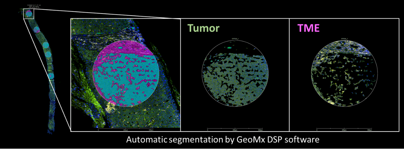
ILL example of segmentation using GeoMX DSP
(c) ILL Department of oncology UNIL CHUV

ILL example of AI-driven segmentation
(c) ILL Department of oncology UNIL CHUV
Biology based segmentation enables near 100% cellularity. Only with spatial profiling we can attribute RNA and protein expression to discrete features of the tissue, mapping region specific biology and cell type abundance. Possibility to profile protein and RNA together on the same slide. GeoMx protocol Tissue integrity is preserved after GeoMx workflow allowing to use slides for downstream analyses as HE staining or IHC.

GeoMx® DSP Human WTA and CTA, Murine WTA, cancer canine atlas are designed to profile all aspects of the tumor and tumor microenvironment biology at RNA level. TCR-profiling might be added to human WTA or CTA to spatially profile the expression of different T cell Receptor variable and joining segments in response to disease onset, progression, treatment, and/or vaccination.

GeoMx® DSP Protein Assays enable quantitative, spatial analysis of up to 570+ proteins from a single FFPE or fresh frozen tissue section, vastly expanding the number of markers you can profile from a single tissue section compared to traditional immunohistochemical methods.
Services provided
|
 |


